EU initiative in digital pathology
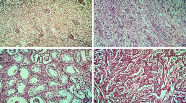
AI is being used increasingly in medicine. To speed development, the EU project BigPicture is building a research platform for digital pathology. It securely collects digital images of tissue samples that are then used to train AI algorithms to interpret the images.
Uppsala University. Photo: Mikael Wallerstedt
“We are creating something that does not actually exist today, a European data warehouse for digital images within pathology,” says Bengt Persson.
Persson is a professor of bioinformatics at Uppsala University and director of National Bioinformatics Infrastructure Sweden (NBIS), which is a platform at SciLifeLab.
The new six-year project within the Innovative Medicines Initiative (IMI) has a budget of 70 million euros. Around 60 partners are participating, both from academia and industry. The focus is on images from pathology, i.e., tissue samples, that are examined under the microscope after being stained in small cross-sections of tissue. As the images are digitised, the collection will grow, opening up new opportunities.
“Instead of interpreting the images using the human eye, machine learning or AI can be used. The advantage of a computer algorithm is that it can look much more carefully, does not get tired like the human eye and can examine millions of images,” says Person.
Requires enormous amounts of data
Training dependable AI applications, however, requires enormous amounts of data, which is a challenge since it requires such large storage capacity and because the images are medical information.
With the NBIS platform, work is now starting to assemble this infrastructure for storage and accessibility of images in collaboration with their Finnish colleagues in Elixir, a European network for life sciences and computer sciences.
“The purpose of this project is to arrange the secure storage of these images. The images are from human data, so legally they are personal data that must be administered securely. This is something we have been working with for four or five years, in terms of sensitive data at the DNA level, in the Federated European Genome-phenome Archive (FEGA) project.”
Technology for sensitive data
The idea is to use the technology already available or in the process of being developed for sensitive DNA data, e.g., to ensure that only authorised individuals can access the data and that the system is secure and protected. At the same time, new components need to be built to process images and compress them.
“The strength of the Swedish side is that we have very good system developers at the NBIS platform. It is primarily their expertise that will be used here.”
What do you see this leading to?
“Making this data available will speed AI development in pathology and that is the goal of all this, to create an infrastructure to train smart algorithms. The idea is that after these six years, this European infrastructure will be made permanent and be used in research and in clinical practice.”
Annica Hulth